

A custom shoulder replacement is a highly specialised technique used in selected cases where the anatomy of the shoulder is significantly altered. This approach involves detailed 3D computer planning to design and manufacture a completely personalised implant tailored to your individual bone structure. The implant is 3D printed in titanium and shaped to fit your shoulder precisely. It is typically considered when there is severe bone loss or distortion, such that standard implants, even when combined with bone grafting, may not provide a stable or reliable reconstruction.
Your shoulder is first assessed through a detailed clinical examination. This includes evaluating:
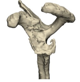
More detailed imaging is required to guide personalised planning and manufacture of the custom component
This may include:
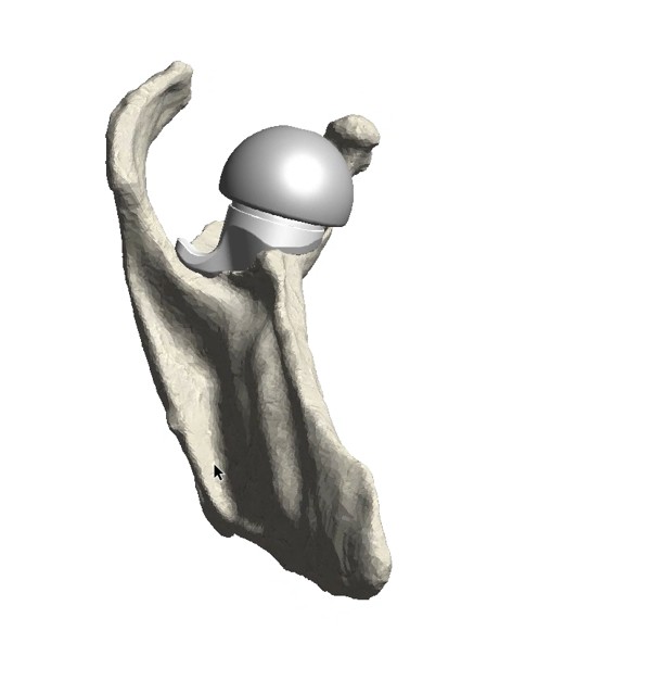
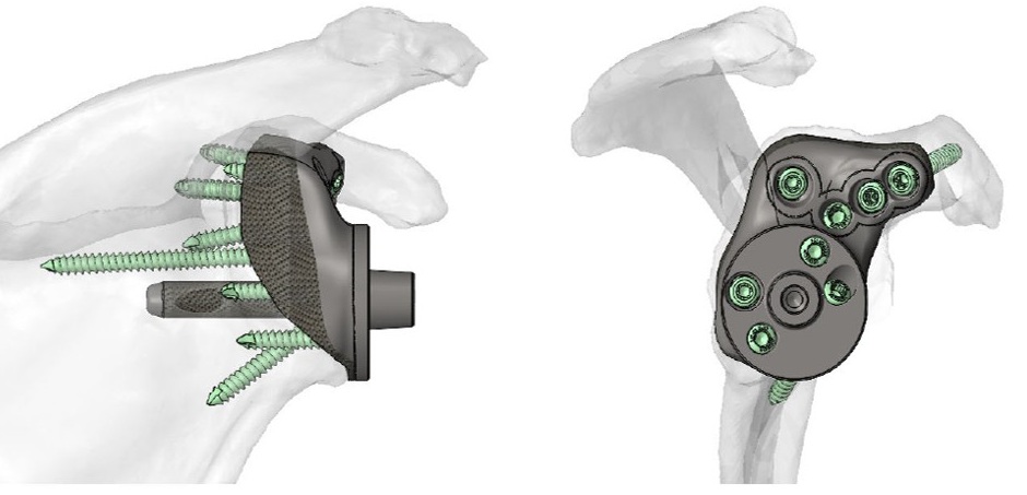
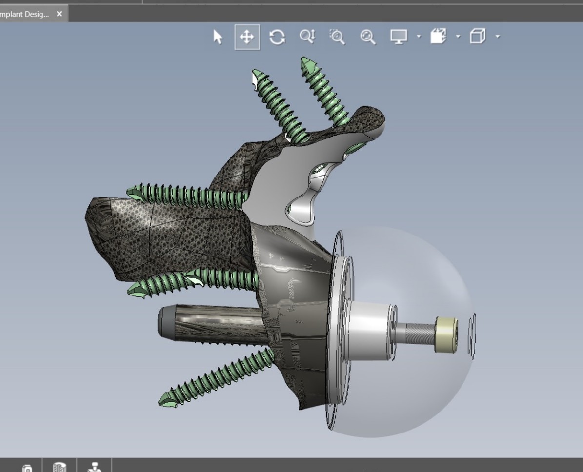
Where a custom implant is required, 3D planning software is used to design a component tailored to your anatomy. Dr Dallalana works with specialised engineers to develop the implant design.
This process involves:
The implantation surgery moves along the same lines as for a reverse shoulder replacement.
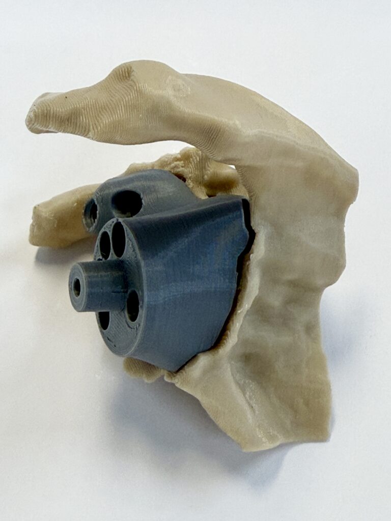
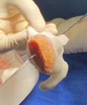
After exposing the shoulder, preparation of the bone (and removal of existing implants if needed) the custom prosthesis is implanted onto the glenoid bone and secured.
The same design software is used to create personalised 3D-printed single-use instruments to perfectly guide the insertion of the custom component.
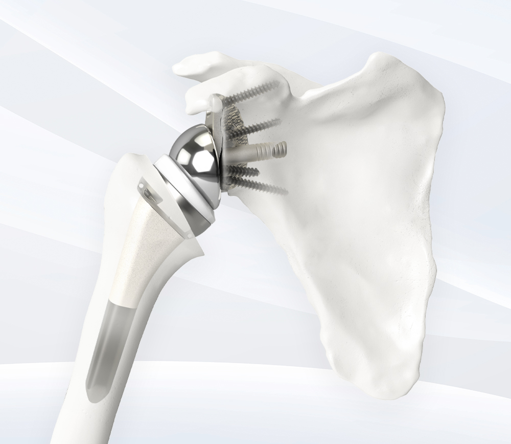
A ball (glenosphere) is then connected to the new custom implant to complete one side of the reverse replacement. A prosthetic stem and socket are placed onto the humerus in a standard way to complete the replacement.
Surgical time is most often longer than standard cases, particularly if it is in the context of a revision procedure.
The same potential complications of surgery exist as for reverse replacement
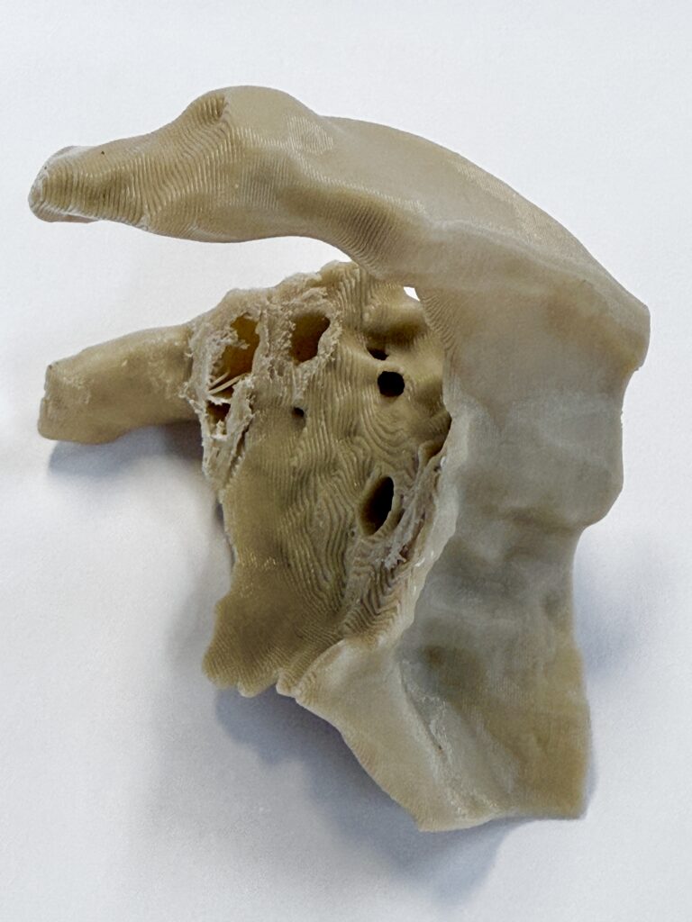
Following your initial assessment, detailed imaging is arranged including a CT scan required for the design software to create the fully customised implant. This can take between 2 and 4 weeks to finalise.
Once the design is finalised, the implant is manufactured using 3D printing technology in titanium.
This process takes time to ensure accuracy and quality. There is typically a lead time of approximately 8 to 10 weeks to allow for production, sterilisation and shipping of the implant before surgery can be scheduled.
In some cases, custom-made implants may involve additional costs, depending on your health fund cover and individual circumstances.
These details are discussed with you in advance so you can make an informed decision. Australian private health funds are not obliged to pay fully for custom implants, and specific requests are made by Dr Dallalana’s office to outline the specific individual circumstances requiring use of the custom solution.
Once the implant has been manufactured and all planning is complete, your surgery is scheduled. Pre-operative medical assessment is routinely arranged to ensure safety of the anaesthetic. No specific exercises or prehabilitation is needed.
Custom shoulder replacement is typically reserved for more complex cases and is not required for most patients undergoing shoulder replacement.
Once healed to bone it serves to function as any standard reverse replacement with a high expectation of pain relief and durability. Range of movement and strength are generally more limited than standard cases due to muscle weakness, scarring and other factors related to complex situations or repeat surgery.
The shoulder in most cases may be used for simple tasks right away after the surgery, as with a standard reverse replacement.
Custom-made implants are designed to address challenges where standard techniques may not be suitable. This approach allows reconstruction to be tailored to your individual anatomy. In many patients it may represent the only viable way to control pain and achieve a functioning shoulder.
Dr Dallalana has a specific interest in complex and revision shoulder replacement and has a high level of experience with custom replacement surgery.
Following surgery, your recovery and rehabilitation pathway is similar to reverse shoulder replacement, with small changes advised individually based on the nature of the custom reconstruction performed.
Length of hospitalisation is similar to that of reverse or revision replacement, 2 days on average.
Message sent